



Company to highlight real-world experience from national screening programs and public healthcare projects
AI applications expand from image analysis to longitudinal comparison, screening results and follow-up management
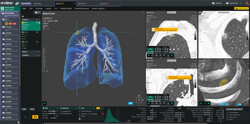
Coreline Soft, a medical imaging artificial intelligence company, will participate in the Korean Congress of Radiology 2026 (KCR 2026), scheduled for Sept. 9-12 at KINTEX in Goyang, South Korea, where it will showcase how chest CT-based AI is being used in clinical practice and population screening programs.
KCR is one of South Korea’s largest radiology conferences, bringing together radiologists, researchers and medical industry professionals from Korea and overseas. Under the theme “AI in Radiology: Beyond Detection,” Coreline Soft will present its key medical imaging AI solutions and real-world applications at Booth B-04.
From detecting lesions to managing patients over time
One of the shifts emerging in medical imaging AI is the move from technology that can simply be deployed to technology that can be used consistently as part of routine clinical workflows.
As AI becomes integrated into medical practice, regulatory approval and accuracy on a single dataset are no longer the only measures of its value. Increasingly important are whether the technology can produce consistent results across hospitals, scanners and readers, connect current scans with previous examinations, quantify changes over time and help ensure that detected abnormalities lead to appropriate follow-up.
Coreline Soft plans to highlight this “post-detection” role of AI at KCR, drawing on its experience with South Korea’s national lung cancer screening program, multi-center public healthcare projects and Germany’s statutory health insurance-based lung cancer screening system.
This is particularly relevant to health checkups and lung cancer screening, where the same individual may undergo CT examinations repeatedly over several years. In these settings, detecting an abnormality on a single scan is only part of the process. Comparing prior images, quantifying changes, assessing risk and managing follow-up examinations are also critical.
Lung cancer screening itself involves a sequence of steps ranging from eligibility assessment and low-dose CT imaging to nodule analysis, risk assessment, reporting and follow-up. As screening programs expand across multiple hospitals and readers, maintaining standardized and consistent workflows becomes increasingly important.
At Booth B-04, Coreline Soft will demonstrate AVIEW LCS Plus, which supports lung nodule detection and quantitative analysis as well as comparison with previous examinations and longitudinal follow-up.
The company will also present how AI-based quantitative analysis is being incorporated into radiology workflows, including its AI technology for lung nodule detection and lung cancer prediction that was recently designated under South Korea’s deferred-assessment pathway for new health technologies.
AVIEW HUB extends this approach to multi-center screening environments. The platform supports connections between readers and participating institutions, data management, quality control and standardized screening workflows, helping AI-generated findings move beyond individual image analysis toward follow-up examinations and subsequent clinical care.
In neuroimaging, Coreline Soft will showcase AVIEW NeuroCAD, which quantitatively analyzes brain atrophy and neurodegenerative changes. Quantitative imaging can provide objective measurements and comparisons with reference populations, supplementing visual assessment that may vary depending on a reader’s experience.
The broader direction is not to replace radiologists’ clinical judgment, but to quantify information contained in medical images and provide additional data that can support clinical decision-making.
One chest CT, multiple health risks
Another trend in imaging AI is extracting more clinically relevant information from a single CT examination.
Beyond pulmonary nodules, AI can analyze multiple findings and risk factors from chest CT scans, including chronic obstructive pulmonary disease (COPD), coronary artery calcification (CAC) and interstitial lung abnormalities (ILA). This expands the potential clinical value of information already contained in a chest CT.
The approach does not mean diagnosing multiple diseases from a single scan. Rather, AI can quantify and structure information relating to the lungs, cardiovascular system and other areas, giving clinicians additional information that may warrant further assessment.
The role of follow-up is also becoming more prominent in national health screening policies. The focus is gradually extending beyond increasing the number of examinations toward helping participants understand their results, identifying high-risk individuals and connecting them with appropriate medical care.
Coreline Soft is pursuing a similar approach by expanding AI-generated information beyond the clinician-facing analysis screen. The company is working to visualize and structure key lung and cardiovascular findings in screening reports that can help participants better understand abnormalities and the possible need for additional examinations.
Germany integrates CAD into screening and follow-up pathways
Coreline Soft’s experience in South Korea’s national lung cancer screening program and public healthcare projects is also being extended to overseas screening markets. Germany, in particular, illustrates how medical imaging AI can become embedded in a broader screening workflow rather than operate solely as a lesion-detection tool.
Germany introduced low-dose CT lung cancer screening for high-risk individuals under its statutory health insurance system in April 2026. The screening framework incorporates computer-aided detection, or CAD, software into the CT interpretation process.
Under the system, an initial reader reviews the CT examination and uses CAD for additional assessment. Findings requiring further evaluation can proceed to an independent second reading, followed by a joint determination by the readers and, when necessary, decisions on subsequent examinations.
The reimbursement structure also extends beyond the initial low-dose CT scan. It includes services associated with follow-up CT examinations within 12 months, referrals for second readings, independent second interpretations, additional consultations and procedures for resolving differing interpretations.
The framework highlights a broader shift in the requirements for medical imaging AI. Detection accuracy remains important, but so do consistency in interpretation, comparison with prior examinations, follow-up management, connections between institutions, quality control and integration with hospital IT systems.
Coreline Soft is addressing this screening environment in Germany with AVIEW LCS, AVIEW LCS Plus and AVIEW HUB. Following the launch of Germany’s LDCT screening program in April, the company had secured 77 new customer accounts as of the end of July, including major institutions such as Charité and Heidelberg University Hospital.
Experience expands across national screening systems
Lung cancer screening models differ considerably by country. While Germany has moved into a reimbursed screening market, Australia is operating its National Lung Cancer Screening Program, or NLCSP. France is developing an organized screening model through the IMPULSION initiative, while Italy is conducting lung cancer early-detection and screening projects through the multi-center RISP network.
Building on its experience with South Korea’s national lung cancer screening program, Coreline Soft has participated in lung cancer screening initiatives across these different healthcare environments, including Australia’s NLCSP, France’s IMPULSION and Italy’s RISP.
Each market has different eligibility criteria, reading protocols and healthcare IT environments. Experience across these programs is therefore increasingly relevant as AI vendors move from supplying standalone detection software toward supporting the operation of large-scale screening systems.
The competitive focus in medical imaging AI is consequently broadening. The question is no longer only whether an algorithm can detect a lesion accurately on a single examination, but whether it can compare previous scans, quantify risk and changes over time, connect patients to follow-up examinations and clinical care, and perform those functions consistently across multiple healthcare institutions.
“The role of AI in radiology is expanding from detecting a single lesion toward quantitative analysis, comparison with prior images, multi-disease analysis, explanation of screening results and follow-up management across the broader healthcare process,” a Coreline Soft official said. “At KCR 2026, we plan to share our real-world experience accumulated in hospitals and public healthcare settings and demonstrate how AI can provide practical support for radiologists.”