



Viz.ai, RapidAI and Brainomix lead in hospital alerts, care coordination and interhospital transfers
Clinical value must include transfer times, thrombectomy speed and functional recovery at 90 days
Series Overview
- September 4 | HIRA Launches First AI-Assisted Claims Review
- September 11 | Medical Imaging AI Platform Competition
- September 18 | Lung Cancer Screening AI and National Follow-Up Care
- September 25 | Breast AI Shows Clinical Value Through Fewer Interval Cancers and Faster Diagnosis
- October 2 | Stroke AI Links Image Analysis to Treatment and Recovery
- October 9 | DICOM Standardization and the Performance and Security Management of Medical Imaging AI
- October 16 | Reimbursement for Medical Imaging AI Based on Clinical Outcomes
- October 23 | Global Market Strategies for Korean Medical Imaging AI
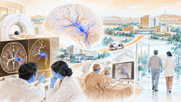
The clinical value of artificial intelligence in stroke imaging must be measured by both diagnostic performance and its effect on treatment times and patient recovery. Rapid identification of an intracranial hemorrhage or large-vessel occlusion produces limited benefit if clinicians see the alert late or if interhospital transfer and mechanical thrombectomy are delayed.
Patients with suspected acute ischemic stroke generally receive non-contrast computed tomography to rule out hemorrhage, followed by CT angiography to identify an occluded artery. CT perfusion imaging or magnetic resonance imaging may then be used to distinguish irreversibly damaged brain tissue from tissue that could still be saved. Once a large-vessel occlusion is confirmed, the hospital must activate a thrombectomy team or transfer the patient to a center capable of performing the procedure.
AI can prioritize emergency scans, identify suspected lesions, calculate their location and volume, and send alerts to clinicians. These products should be evaluated across four separate areas: image analysis, regulatory clearance, clinical workflow integration and patient outcomes. Regulatory approvals and hospital installations show market access, but they do not establish faster treatment or better functional recovery at 90 days.
A research team led by Jeffrey Saver, a professor at the University of California, Los Angeles, analyzed 1,287 patients enrolled in five randomized trials. The study found that the benefit of thrombectomy declined as the time between symptom onset and treatment increased.
When treatment began three hours after symptom onset, the absolute difference in the likelihood of achieving a lower disability score was 39.2%. That figure fell to 30.2% at six hours and 15.7% at eight hours. Stroke AI must therefore be assessed by how much it accelerates reperfusion treatment, as well as by how quickly it processes an image.
JLK and Global Rivals Require the Same Evaluation Standards
South Korea’s JLK has assembled 12 stroke imaging applications under its MEDIHUB STROKE portfolio. Its products analyze CT, CT angiography, CT perfusion and MRI scans for conditions including intracranial hemorrhage, large-vessel occlusion and acute cerebral infarction.
According to JLK’s business report filed in March 2026, JLK-LVO, JLK-CTP, JLK-PWI, JLK-ICH and JLK-SDH have received 510(k) clearance from the US Food and Drug Administration. Seven products, including JLK-DWI, have passed Japan’s medical-device review process. In South Korea, JLK-DWI, which analyzes ischemic lesions on diffusion-weighted MRI, has entered the non-reimbursed clinical pathway for innovative medical technologies.
These clearances permit the company to market products in the United States and Japan. FDA 510(k) clearance establishes substantial equivalence to a legally marketed device; it does not determine that the new product is clinically superior to competing systems. Multiple product approvals also do not demonstrate that the applications operate together as one integrated hospital workflow.
JLK’s image-analysis performance has been examined in South Korean studies. In a multicenter study involving 595 patients, JLK-LVO achieved sensitivity of 85.8% and specificity of 96.9% in detecting large-vessel occlusion. Less-experienced physicians also improved their detection sensitivity when using the AI system.
A 2026 South Korean real-world comparison analyzed images from 176 patients with suspected stroke using JLK-LVO and RAPID CTA from US-based RapidAI. Both systems achieved an area under the receiver operating characteristic curve of 0.93. At a matched sensitivity of 83%, specificity was 96% for JLK-LVO and 89% for RAPID CTA.
The results do not establish that JLK-LVO is generally superior to RAPID CTA. The study was a retrospective analysis conducted at a single stroke center, and a JLK researcher was among the co-authors. Both systems sometimes classified severe arterial stenosis as an occlusion and missed distal occlusions when collateral circulation was well developed.
Another study compared JLK-DWI and RAPID MRI with manual lesion measurements in diffusion-weighted MRI scans from 414 patients. RAPID MRI failed to detect lesions in 61.4% of cases, compared with 1.9% for JLK-DWI. Infarct volumes calculated by both systems correlated strongly with manual measurements.
JLK-DWI performed better in detecting small lesions in this study. The research, however, compared specific versions of the two products and included participation by JLK personnel. It did not determine whether detecting more lesions changed decisions on thrombolysis or thrombectomy or improved functional recovery at 90 days.
A separate CT perfusion study illustrates why hospitals should not use AI-generated measurements as the sole basis for treatment decisions. Researchers compared JLK-CTP and RAPID in 327 South Korean patients and found differences in the volumes calculated for the ischemic core and hypoperfused tissue.
Both systems tended to overestimate the volume of irreversibly damaged tissue and showed weaker agreement with early follow-up MRI in patients with small infarcts. An overestimated ischemic core could exclude a potentially eligible patient from thrombectomy. Clinicians must therefore examine the original images, neurological findings and time of symptom onset alongside the AI-generated measurements.
JLK-NCCT, which received US clearance in 2026, analyzes non-contrast CT scans for suspected intracranial hemorrhage and large-vessel occlusion and sends mobile alerts. FDA documentation defines it as a prioritization and notification tool rather than a diagnostic device. A negative result cannot rule out disease, and standard care may still require CT angiography or MR angiography.
JLK has established a broad imaging portfolio, regulatory clearances in the United States and Japan, and several direct product comparisons. Its published research, however, remains concentrated on lesion detection and volume measurement. Publicly available multicenter evidence remains limited on overseas use, clinician adoption, alert-response times, transfer times, repeat imaging, thrombectomy initiation and functional recovery at 90 days.
US-based Viz.ai detects suspected large-vessel occlusions, sends mobile alerts and allows referring hospitals and thrombectomy centers to share images and patient information. RapidAI, also based in the United States, combines occlusion detection and perfusion analysis with transfer decisions. UK-based Brainomix supplies non-contrast CT, CT angiography and CT perfusion products to healthcare providers across Europe.
JLK offers a broad range of CT and MRI applications. Viz.ai and RapidAI have accumulated more evidence on mobile collaboration, care coordination and interhospital transfers, while Brainomix has expanded its experience within European health systems and national programs. JLK has developed imaging technology and regulatory access that allow comparison with global competitors, but it has released less evidence on overseas hospital operations and patient outcomes.
Faster Treatment Must Lead to Better Recovery at 90 Days
A 2026 study by researchers at the US Methodist Health System reviewed 12 studies of AI-based stroke triage and notification systems. Published in Cardiovascular and Interventional Radiology, the journal of the Cardiovascular and Interventional Radiological Society of Europe, it found that AI implementation was associated with a 17.12-minute reduction in the time from hospital arrival to groin puncture for thrombectomy.
The time to the first thrombectomy pass fell by 26.55 minutes, while the time to revascularization declined by 14.55 minutes. For patients transferred from another hospital, the time between arrival and departure from the referring hospital was shortened by 36.8 minutes.
Only one of the 12 studies was a clinical trial; the remaining 11 were observational studies. The researchers concluded that randomized controlled trials were still needed. A separate published review also found shorter times to groin puncture and reperfusion after AI implementation but no statistically significant improvement in functional independence at 90 days or mortality.
Most real-world studies compare performance before and after an AI system is introduced. Hospitals may revise emergency procedures, imaging protocols, transfer rules and staff training during the same period. This makes it difficult to separate the effect of the algorithm from broader operational improvements.
A prospective multicenter study in Switzerland found that AI implementation reduced the time from imaging to a transfer request by about seven minutes and shortened the interval from emergency department arrival to groin puncture by approximately 31 minutes. The proportion of patients receiving CT angiography at referring hospitals rose from 28% to 81%, while repeat imaging at receiving centers fell from 79% to 42%.
The study’s primary endpoint—the time from patient arrival to the transfer request—did not improve significantly. The findings therefore do not show that AI alone accelerated the transfer decision. Wider use of CT angiography, fewer repeat scans and changes in interhospital workflow also contributed to faster treatment.
Tessa Cook, a professor of radiology at the University of Pennsylvania and chair of the committee that developed the first imaging AI practice parameter from the American College of Radiology and the Society for Imaging Informatics in Medicine, said the guidance covers the entire process from selecting an AI product to performance monitoring and continuous quality improvement. She has emphasized that hospitals must continuously manage the deployment, use and updating of rapidly changing clinical AI.
The same standards should apply to JLK and its overseas competitors. Performance can vary across hospitals as patient populations, scanners and imaging protocols change. Healthcare providers should test products on local data before deployment and continuously monitor false positives, false negatives, processing times and performance changes following software updates.
In South Korea, the effectiveness of stroke AI will depend on coordination between regional hospitals and the country’s cardiovascular and cerebrovascular disease centers. An alert from a regional hospital will not accelerate treatment if an ambulance, receiving bed or thrombectomy team is unavailable. Hospitals should report alert-response times, transfer-acceptance times, repeat imaging rates and thrombectomy rates, rather than relying only on installation numbers and scan volumes.
Patient recovery can be measured using the modified Rankin Scale, or mRS, 90 days after a stroke. The scale grades disability from 0 to 6, with 0 indicating no symptoms and 6 representing death. Scores of 0 to 2 generally indicate functional independence in daily life.
JLK has built an imaging portfolio and diagnostic performance that can be compared with leading international companies. Its publicly available evidence, however, remains focused on regulatory clearances in the United States and Japan and diagnostic studies conducted primarily in South Korea. Independent multicenter research on overseas clinical use, interhospital transfers, treatment times and functional recovery at 90 days remains insufficient to establish its clinical competitiveness in global markets.
Viz.ai, RapidAI and Brainomix have published more real-world evidence on lesion detection, clinician alerts and treatment-time reductions than JLK. However, evidence that their faster workflows consistently improve functional recovery, mortality and healthcare costs across multiple independent institutions also remains limited.
Domestic and international companies should therefore be evaluated under the same measures: the number of active clinical sites, interhospital transfer times, treatment speed, functional recovery at 90 days, mortality and changes in medical costs. These measures distinguish product deployment from demonstrated clinical value.
The role of stroke AI does not end when it marks an occluded artery. Its clinical value is established when alerts accelerate specialist review, interhospital transfer and thrombectomy and reduce long-term disability. The international competitiveness of South Korean medical imaging AI companies will depend on their ability to demonstrate this entire process through independent real-world evidence.
Part 6 will examine how the DICOM standard for medical-image descriptions and metadata enables hospitals to operate multiple AI applications safely.